")
5-2. Lesson 5 (cont) Supraventricular Arrhythmias
Topics of Study:
- Premature atrial complexes
- Premature junctional complexes
- Atrial fibrillation
- Atrial flutter
- Ectopic atrial tachycardia and rhythm
- Multifocal atrial tachycardia
- Paroxysmal supraventricular tachycardia
- Junctional rhythms and tachycardias
Premature atrial complexes
- Occur as single or repetitive events and have unifocal or multifocal origins.
- The ectopic P wave (called P') is often hidden in the ST-T wave of the preceding beat. (Dr. Marriott, master ECG teacher and author, likes to say: "Cherchez le P on let T" which in French means: "Search for the P on the T wave", but it's more sexy in French!)
- The P'R interval is normal or prolonged because the AV junction is often partially refractory when the premature impulse enters it.
- PAC's can have three different outcomes depending on the degree of prematurity (i.e., coupling interval from previous P wave), and the preceding cycle length. This is illustrated in the "ladder" diagram where normal sinus beats (P) are followed by three possible PACs; in the diagram the refractory periods of the AV node and bundle branches are indicated by the width of the boxes:
- Outcome #1. Nonconducted (blocked); i.e., no QRS complex because the PAC finds AV node still refractory. (see PAC labeled 'a' in the upper diagram 1)
- Outcome #2. Conducted with aberration; i.e., PAC makes it into the ventricles but finds one or more of the conducting fascicles or bundle branches refractory. The resulting QRS is usually wide, and is sometimes called an Ashman beat (see PAC 'b' in diagram 1)
- Outcome #3. Normal conduction; i.e., similar to other QRS complexes in the ECG. (See PAC 'c' in the diagram 1)
- In the diagram 2, seen above, the cycle length (i.e., PP interval) has increased (slower heart rate), and this results in increased refractoriness of all the structures in the conduction system (i.e., wider boxes). PAC 'b' now can't get through the AV node and is nonconducted; PAC 'c' is now blocked in the right bundle branch and results in a RBBB QRS complex (aberrant conduction); PAC 'd' is far enough away to conduct normally. Therefore, the fate of a PAC depends on 1) the coupling interval from the last P wave and 2) the preceding cycle length or heart rate.
- The pause after a PAC is usually incomplete; i.e., the PAC usually enters the sinus node and resets its timing, causing the next sinus P to appear earlier than expected. (PVCs, on the other hand, are usually followed by a complete pause because the PVC does not usually perturb the sinus node; see ECG below.)


Premature junctional complexes
- Similar to PAC's in clinical implications, but occur less frequently.
- The PJC focus, located in the AV junction, captures the atria (retrograde) and the ventricles (antegrade). The retrograde P wave may appear before, during, or after the QRS complex; if before, the PR interval is usually short (i.e., < 0.12 s). The ECG tracing and ladder diagram shown below illustrates two classic PJC's with retrograde P waves following the QRS.

Atrial Fibrillation (A-fib)

- Atrial activity is poorly defined; may see course or fine undulations or no atrial activity at all. If atrial activity is seen, it resembles an old saw (when compared to atrial flutter that often resembles a new saw).
- Ventricular response is irregularly irregular and may be fast (HR >100 bpm, indicates inadequate rate control), moderate (HR = 60-100 bpm), or slow (HR < 60 bpm, indicates excessive rate control, AV node disease, or drug toxicity).
- A regular ventricular response with A-fib usually indicates complete AV block with an escape or accelerated ectopic pacemaker originating in the AV junction or ventricles (i.e., must consider digoxin toxicity or AV node disease).
- The differential diagnosis includes atrial flutter with an irregular ventricular response and multifocal atrial tachycardia (MAT), which is usually irregularly irregular. The differential diagnosis may be hard to make from a single lead rhythm strip; the 12-lead ECG is best for differentiating these three arrhythmias.
Atrial Flutter (A-flutter):

- Regular atrial activity with a "clean" saw-tooth appearance in leads II, III, aVF, and usually discrete 'P' waves in lead V1. The atrial rate is usually about 300/min, but may be as slow as 150-200/min or as fast as 400-450/min.
- Untreated A-flutter often presents with a 2:1 A-V conduction ratio. This is the most commonly missed supraventricular tachycardia because the flutter waves are often difficult to find when there is 2:1 ratio. Therefore, always think "atrial flutter with 2:1 block" whenever there is a regular supraventricular tachycardia @ ~150 bpm! (You won't miss it if you look for it in a 12-lead ECG)
- The ventricular response may be 2:1, 3:1 (rare), 4:1, or irregular depending upon the AV conduction properties and AV node slowing drugs on board (e.g., digoxin, beta blockers).
Ectopic Atrial Tachycardia and Rhythm
- Ectopic, discrete looking, unifocal P' waves with atrial rate < 250/min (not to be confused with slow atrial flutter)
- Ectopic P' waves usually precede QRS complexes with P'R interval < RP' interval (i.e., not to be confused with paroxysmal supraventricular tachycardia with retrograde P waves appearing shortly after the QRS complexes).
- Ventricular response may be 1:1 or with varying degrees of AV block (especially in digitalis toxicity, as shown in this 3-lead ECG with 2:1 block).
- Ectopic atrial rhythm is similar to ectopic atrial tachycardia, but with HR < 100 bpm.

Multifocal Atrial Tachycardia (MAT) and rhythm
- Discrete, multifocal P' waves occurring at rates of 100-250/min and with varying P'R intervals (should see at least 3 different P wave morphologies in a given lead).
- Ventricular response is irregularly irregular (i.e., often confused with A-fib).
- May be intermittent, alternating with periods of normal sinus rhythm.
- Seen most often in elderly patients with chronic or acute medical problems such as exacerbation of chronic obstructive pulmonary disease.
- If atrial rate is < 100 bpm, call it multifocal atrial rhythm
Paroxysmal Supraventricular Tachycardia (PSVT)
- Basic Considerations: These arrhythmias are circus movement or reciprocating tachycardias because they utilize the mechanism of reentry. The onset is sudden, usually initiated by a premature beat, and the arrhythmia also stops abruptly - which is why they are called paroxysmal. They are usually narrow-QRS tachycardias unless there is preexisting bundle branch block or rate-related aberrant ventricular conduction. There are several types of PSVT depending on the location of the reentry circuit.
- AV Nodal Reentrant Tachycardia (AVNRT): This is the most common form of PSVT accounting for approximately 50% of all symptomatic PSVTs. The diagram illustrates the probable mechanism involving dual AV nodal pathways, alpha and beta, with different electrical properties. In the diagram alpha is a fast AV nodal pathway with a long refractory period (RP), and beta is the slow pathway with a short RP. During sinus rhythm alpha is always used because it conducts faster. An early PAC, however, finds alpha still refractory and must use the slower beta pathway to reach the ventricles. By the time it traverses beta, however, alpha has recovered allowing retrograde conduction back to the atria. The retrograde P wave (called an atrial echo for obvious reasons) is often simultaneous with the QRS and, therefore, not seen on the ECG, but it can reenter the AV junction because of beta's short RP.
- AV Reciprocating Tachycardia (Extranodal bypass pathway): This is the second most common form of PSVT and is seen in patients with WPW syndrome. The WPW ECG, seen in the diagram, shows a short PR, delta wave, and somewhat widened QRS.
- Sino-Atrial Reentrant Tachycardia: This is a rare form of PSVT where the reentrant circuit is between the sinus node and the right atria. The ECG looks like sinus tachycardia, but the tachycardia is paroxysmal; i.e., it starts and ends abruptly.


Junctional Rhythms and Tachycardias
Junctional Escape Beats
These are passive, protective beats originating from subsidiary pacemaker cells in the AV junction (usually in the Bundle of His). The pacemaker's basic firing rate is 40-60 bpm; junctional escapes are protective events that occur whenever the primary pacemaker (i.e., sinus node) defaults or the AV node blocks the atrial impulse. The ECG strip shows intermittent sinus slowing with two junctional escapes.

Junctional Escape Rhythm
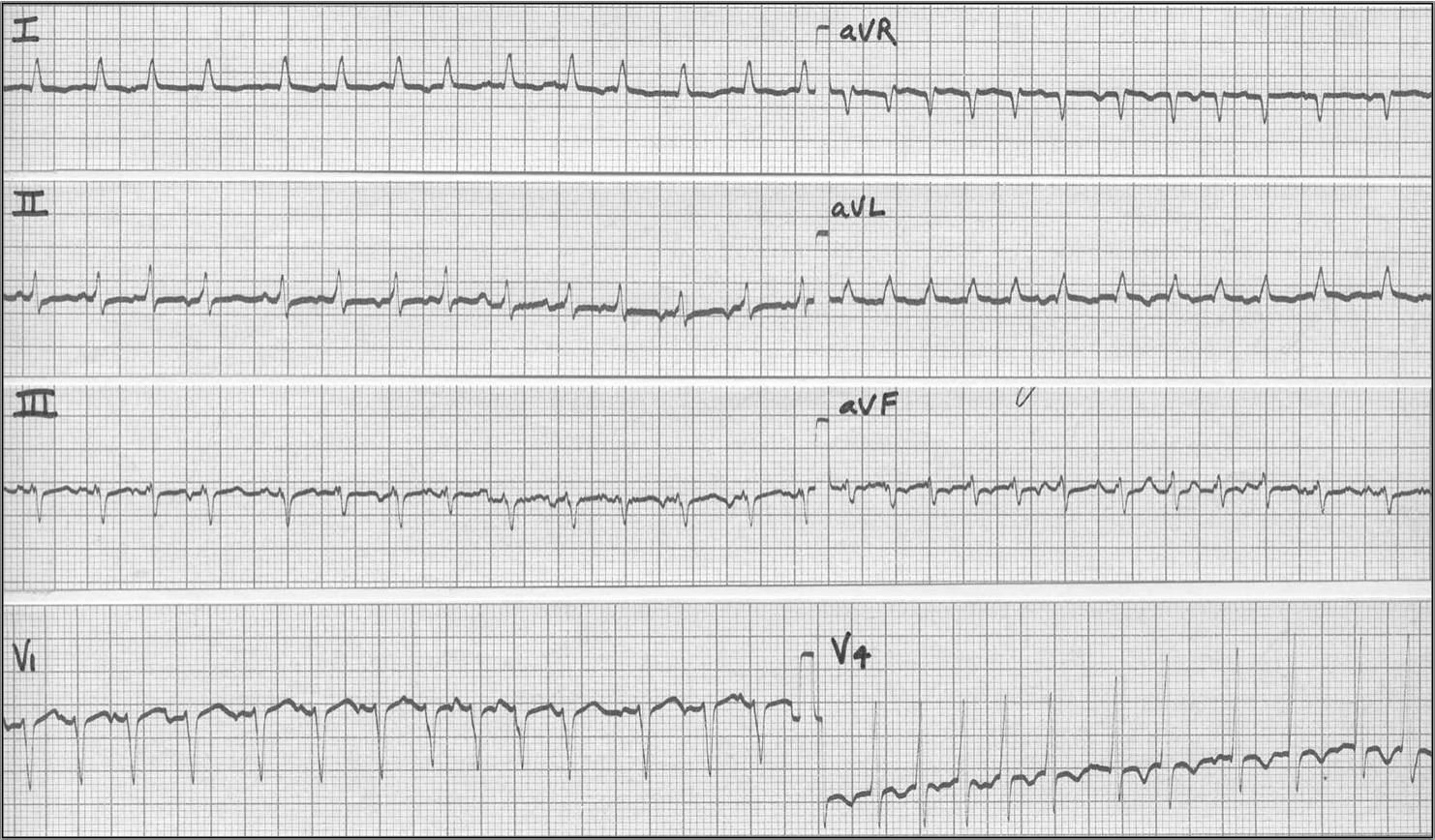
A Junctional Escape Rhythm is a sequence of 3 or more junctional escapes occurring by default at a rate of 40-60 bpm. The 12-lead ECG shown below illustrates a junctional escape rhythm in a well-trained athlete whose resting sinus rate is slower than the junctional rate. Retrograde P waves are hidden in the ST-T waves and best seen in leads II, III, and aVF.

Accelerated Junctional Rhythm
This is an active junctional pacemaker rhythm caused by events that perturb pacemaker cells (e.g., ischemia, drugs, and electrolyte abnormalities). The rate is 60-100 bpm).

Nonparoxysmal Junctional Tachycardia
This usually begins as an accelerated junctional rhythm but the heart rate gradually increases to > 100 bpm. There may be AV dissociation, or retrograde atrial capture may occur. Ischemia (usually from right coronary artery occlusion) and digitalis intoxication are the two most common causes. In the example below junctional tachycardia is seen with ('B') and without exit block ('A').
