")
2-1. How to Measure the QRS Axis
Topics for study:
Introduction
The frontal plane QRS axis represents only the average direction of ventricular activation in the frontal plane. As such this measure can inform the ECG reader of changes in the sequence of ventricular activation (e.g., left anterior fascicular block), or it can be an indicator of myocardial damage (e.g., inferior myocardial infarction).
In the diagram below the normal range is identified (-30° to +90°). Left axis deviation (i.e., superior and leftward) is defined from -30° to -90°, and right axis deviation (i.e., inferior and rightward) is defined from +90° to +150°.
Click to see causes of abnormal axis (lesson 4).
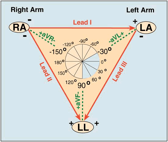
QRS Axis Determination
- First find the isoelectric lead if there is one; i.e., the lead with equal forces in the positive and negative direction. Often this is the lead with the smallest QRS.
- The QRS axis is perpendicular to that lead's orientation (see above diagram).
- Since there are two perpendiculars to each isoelectric lead, chose the perpendicular that best fits the direction of the other ECG leads.
- If there is no isoelectric lead, there are usually two leads that are nearly isoelectric, and these are always 30° apart. Find the perpendiculars for each lead and chose an approximate QRS axis within the 30° range.
- Occasionally each of the 6 frontal plane leads is small and/or isoelectric. The axis cannot be determined and is called indeterminate. This is a normal variant.
Examples of QRS Axis
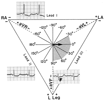
Axis in the normal range:
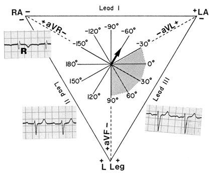
Axis in the left axis deviation (LAD) range:
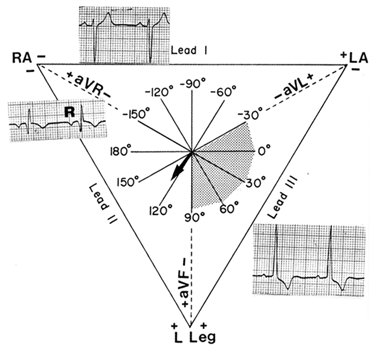
Axis in the right axis deviation (RAD) range: