")
10. ST Segment Abnormalities
Topics for study:
General Introduction to ST, T, and U wave abnormalities
Basic Concept: the specificity of ST-T and U wave abnormalities is provided more by the clinical circumstances in which the ECG changes are found than by the particular changes themselves. Thus the term, nonspecific ST-T wave abnormalities, is frequently used when the clinical data are not available to correlate with the ECG findings. This does not mean that the ECG changes are unimportant! It is the responsibility of the clinician providing care for the patient to ascertain the importance of the ECG findings.
Factors affecting the ST-T and U wave configuration include:
- Intrinsic myocardial disease (e.g., myocarditis, ischemia, infarction, infiltrative or myopathic processes)
- Drugs (e.g., digoxin, quinidine, tricyclics, and many others)
- Electrolyte abnormalities of potassium, magnesium, calcium
- Neurogenic factors (e.g., stroke, hemorrhage, trauma, tumor, etc.)
- Metabolic factors (e.g., hypoglycemia, hyperventilation)
- Atrial repolarization (e.g., at fast heart rates the atrial T wave may pull down the beginning of the ST segment)
- Ventricular conduction abnormalities and rhythms originating in the ventricles
"Secondary" ST-T Wave changes (these are normal ST-T wave changes solely due to alterations in the sequence of ventricular activation):
- ST-T changes seen in bundle branch blocks (generally the ST-T polarity is opposite to the major or terminal deflection of the QRS)
- ST-T changes seen in fascicular block
- ST-T changes seen in nonspecific IVCD
- ST-T changes seen in WPW preexcitation
- ST-T changes in PVCs, ventricular arrhythmias, and ventricular paced beats
"Primary" ST-T Wave Abnormalities (ST-T wave changes that are independent of changes in ventricular activation and that may be the result of global or segmental pathologic processes that affect ventricular repolarization):
- Drug effects (e.g., digoxin, quinidine, etc)
- Electrolyte abnormalities (e.g., hypokalemia)
- Ischemia, infarction, inflammation, etc
- Neurogenic effects (e.g., subarrachnoid hemorrhage causing long QT)
Differential Diagnosis of ST Segment Elevation
Normal Variant "Early Repolarization" (usually concave upwards, ending with symmetrical, large, upright T waves)
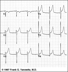
Example #1: "Early Repolarization": note high take off of the ST segment in leads V4-6; the ST elevation in V2-3 is generally seen in most normal ECG's; the ST elevation in V2-6 is concave upwards, another characteristic of this normal variant.
Ischemic Heart Disease (usually convex upwards, or straightened)
- Acute transmural injury - as in this acute anterior MI
- Persistent ST elevation after acute MI suggests ventricular aneurysm
- ST elevation may also be seen as a manifestation of Prinzmetal's (variant) angina (coronary artery spasm)
- ST elevation during exercise testing suggests extremely tight coronary artery stenosis or spasm (transmural ischemia)

Acute Pericarditis
- Concave upwards ST elevation in most leads except aVR
- No reciprocal ST segment depression (except in aVR)
- Unlike "early repolarization", T waves are usually low amplitude, and heart rate is usually increased.
- May see PR segment depression, a manifestation of atrial injury
Other Causes:
- Left ventricular hypertrophy (in right precordial leads with large S-waves)
- Left bundle branch block (in right precordial leads with large S-waves)
- Advanced hyperkalemia
- Hypothermia (prominent J-waves or Osborne waves)
Differential Diagnosis of ST Segment Depression
Normal variants or artifacts:
- Pseudo-ST-depression (wandering baseline due to poor skin-electrode contact)
- Physiologic J-junctional depression with sinus tachycardia (most likely due to atrial repolarization)
- Hyperventilation-induced ST segment depression
- Ischemic heart disease
- Subendocardial ischemia (exercise induced or during angina attack - as illustrated below)
- ST segment depression is often characterized as "horizontal", "upsloping", or "downsloping"
- ST segment depression is often characterized as "horizontal", "upsloping", or "downsloping"
- Non Q-wave MI
- Reciprocal changes in acute Q-wave MI (e.g., ST depression in leads I & aVL with acute inferior MI)
- Subendocardial ischemia (exercise induced or during angina attack - as illustrated below)
- Nonischemic causes of ST depression
- RVH (right precordial leads) or LVH (left precordial leads, I, aVL)
- Digoxin effect on ECG
- Hypokalemia
- Mitral valve prolapse (some cases)
- CNS disease
- Secondary ST segment changes with IV conduction abnormalities (e.g., RBBB, LBBB, WPW, etc)

