")
ECG Image Index
The following ECG categories contain hundreds of ECGs that range from the sublime to the ridiculous, from simplicity to complexity, and from boring to fascinating. Many of the ECG rhythm strips come from the collection of the late Dr. Alan Lindsay, master teacher of electrocardiography. Most of the 12- and 6-lead ECGs were recorded at LDS Hospital in Salt Lake City, Utah. Marquette Electronics has also given permission to use ECG rhythms and diagrams from their educational posters. Each of the ECGs has an interpretation and many have additional explanations that help explain the diagnosis. Feedback is encouraged using the feedback form provided with this website.
ECG Categories
- Diagrams & Pictures
- Frontal Plane QRS Axis Examples
- Sinus, Atrial, Junctional Rhythms
- PACs, PJCs, and PVCs
- Atrial Fibrillation, Flutter and SVT's
- Ventricular Rhythms and Tachycardias
- Mischief in the AV Junction (Blocks and Dissociation)
- Bundle Branch, Fascicular Blocks, and WPW
- Artificial Pacemakers
- Myocardial Infarctions
- Hypertrophies and Enlargements
- ST-T and U Wave Abnormalities and Long QT
- Odds & Ends
Diagrams & Pictures
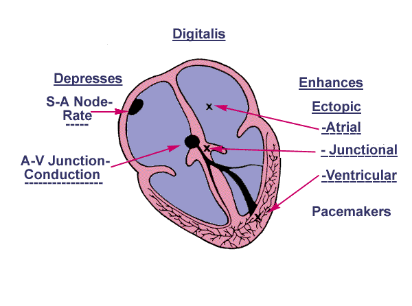
- Diagram: Digitalis Effect on Rhythm and Conduction-KH
- WPW Diagram-KH
- ECG Intervals and Waves-KH
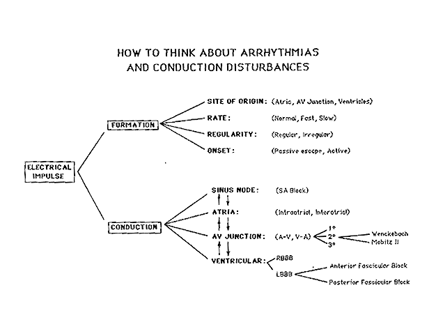
- Conceptual Framework: Arrhythmias and Conduction Abnormalities-KH
- Cardiac Conduction System Diagram
- Compensatory vs. Non-compensatory Pauses
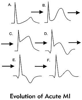
- Diagram: Stages of Acute Q-Wave MI-KH
- Alan E. Lindsay, MD: A Teacher of Substance and Style
- Dr. Alan E. Lindsay
- All About Premature Beats-KH
- The Three Fates Of PAC's: 1. Normal Conduction; 2. Aberrant Conduction; 3. Non-conduction-KH
- Diagram: AV Nodal Reentrant Tachycardia-KH
- Diagram: Type I vs. Type II 2nd Degree AV Block-KH
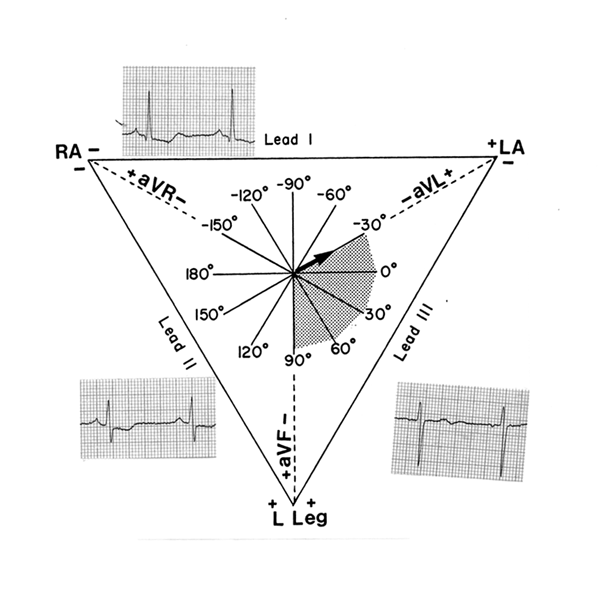
- Diagram: Frontal Plane Leads-KH
- ST Segment Depression Morphologies
- Frontal and Horizontal Plane Lead Diagram-KH
- Differential Dx of upgoing wide QRS's in V1
- Differential Dx of downgoing wide QRS's in V1
- Differential Dx of downgoing wide QRS's in V1
- ECG Ischemic Changes with Excersize
- Locations of Conduction Abnormalities
Frontal Plane QRS Axis Examples
- QRS Axis = +90 degrees-KH
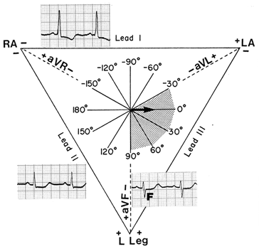
- QRS Axis = -30 degrees-KH
- QRS Axis = 0 degrees-KH
- Left Axis Deviation: QRS Axis = -60 degrees-KH
- QRS Axis = +60 degrees-KH
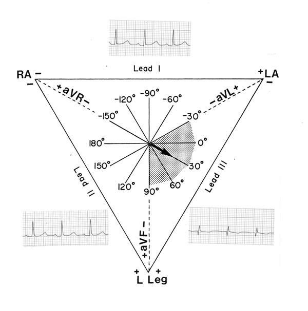
- QRS Axis = +30 degrees-KH
- Left Axis Deviation: QRS Axis = -45 degrees-KH
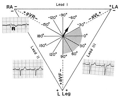
- Right Axis Deviation: QRS Axis = +130 degrees-KH
- Frontal Plane QRS Axis = +90 degrees-KH
- Frontal Plane QRS Axis = +75 degrees-KH
- Frontal Plane QRS Axis = +50 degrees-KH
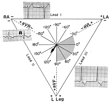
- Frontal Plane QRS Axis = +150 degrees (RAD)-KH
- Frontal Plane QRS Axis = 90 degrees-KH
- Frontal Plane QRS Axis = +30 degrees-KH
- Frontal Plane QRS Axis = +15 degrees-KH
- Frontal Plane QRS Axis = 0 degrees-KH
- Frontal Plane QRS Axis = -15 degrees-KH
- Frontal Plane QRS Axis = -45 degrees-KH
- Frontal Plane QRS Axis = -45 degrees-KH
- Frontal Plane QRS Axis = -75 degrees-KH
- Indeterminate Frontal Plane QRS Axis-KH
- Right Axis Deviation
- Left Axis Deviation
Sinus, Atrial, Junctional Rhythms
- Normal 12 Lead ECG
- Wandering Atrial Pacemaker-KH
- Accelerated Junctional Rhythm
- Sinus Pause with sinus arrhythmia
- Sinus arrhythmia and accelerated ventricular rhythm
- Junctional Escape Rhythm
- Sinus bradycardia with RBBB (HR 38 bpm)
- Accelerated J Rhythm
- Accelerated J Rhythm and 3rd degree AVB
- Accelerated J Rhythm and Incomplete AV Diss
- Normal ECG
- SA block and RBBB
- Sinus Tachycardia
- Sino-Atrial Exit Block Type I
- Sino-Atrial Exit Block Type II
PACs, PJCs, and PVCs
- PAC With RBBB aberration
- What are those funny looking beats????
- Long QT Mischief
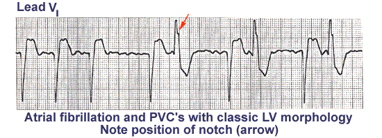
- Left Ventricular PVC's
- Atrial Parasystole
- Ventricular Parasystole
- Ventricular Fusion Beats
- PVC With Venticular Echo
- Nonconducted PACs and Junctional Escapes
- Nonconducted And Conducted PAC's
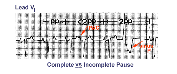
- PAC and PVC: Complete vs. Incomplete Pause-KH
- Identification of PVC's and PAC's-KH
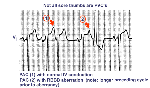
- Not All Sore Thumbs Are Ventricular In Origin-KH
- Nonconducted PAC's: An Unusual Bigeminy-KH
- An Interpolated PAC-KH
- PAC's With RBBB Aberration-KH
- The Three Fates Of PAC's-KH
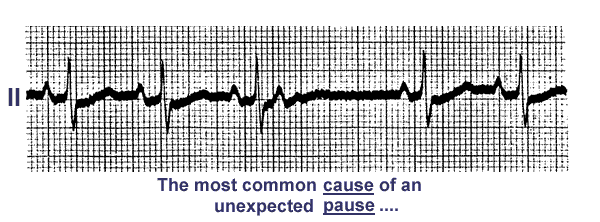
- A Nonconducted PAC Causes An Unexpected Pause-KH
- Nonconducted PAC's Slowing The Heart Rate-KH
- Atrial Parasystole-KH
- Atrial Parasystole-KH
- Nonconducted and Aberrantly Conducted PAC's-KH
- Sore Thumbs-KH
- Junctional Parasystole and Pseudo-AV Block-KH
- Premature Junctional Complexes With Retrograde P Waves-KH
- PAC's With and Without Aberrant Conduction
- Ventricular Bigeminy
- Nonconducted PAC
- PVC Couplets And Triplets
- Interpolated PVC's
- Multifocal PVCs
- Unifocal PVC’s In A Pattern Of Bigeminy
Atrial Fibrillation, Flutter and SVT's
- Atrial Flutter With 2:1 AV Conduction-KH
- Atrial Flutter With 2:1 Conduction: Leads II, III, V1-KH
- Atrial Flutter With 3:2 AV Conduction: 12 lead ECG
- Atrial Flutter with 3:2 Conduction Ratio: lead V1
- Atrial Flutter With 2:1 AV Conduction-KH
- Atrial Flutter With 2:1 AV Conduction: Lead V1-KH
- Atrial Flutter With 2:1 AV Conduction-KH
- Atrial Flutter With 2:1 AV Conduction: Leads II, III, V1-KH
- LBBB and Atrial Flutter with 2:1 AV Block
- Multifocal Atrial Tachycardia (MAT)
- Atrial Fibrillation in Patient with WPW Syndrome
- A PAC Initiates Paroxsymal Atrial Fibrillation-KH
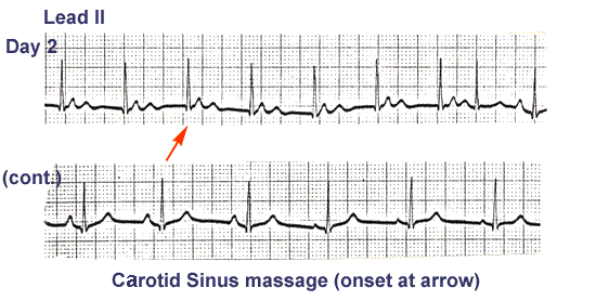
- Massage Parlor Games-KH
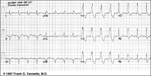
- Atrial Flutter With Variable AV Block And Rate-Dependent LBBB-KH
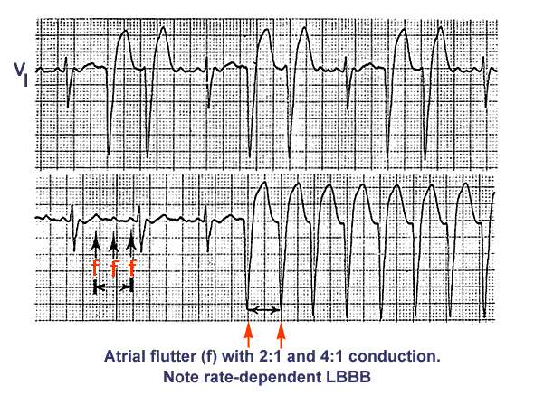
- Atrial Flutter With 2:1 and 4:1 Conduction and Rate Dependent LBBB-KH
- Atrial tachycardia With 3:2 AV Block-KH
- Atrial Tachycardia With 3:2 and 2:1 AV Block-KH
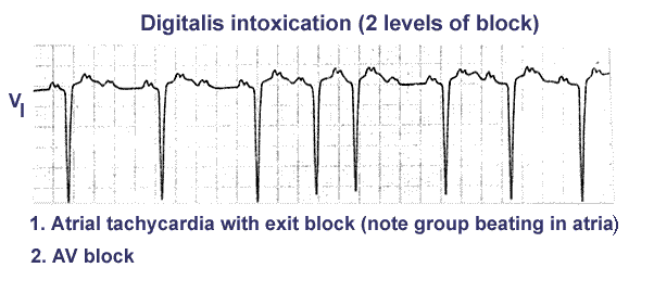
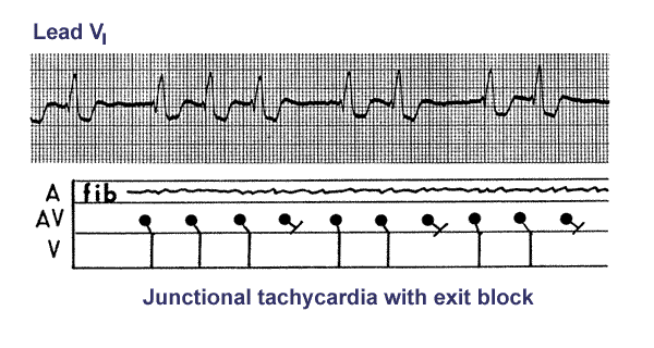
- Digitalis Intoxication: Junctional Tachycardia With and Without AV Block-KH
- Digitalis Intoxication: Junctional Tachycardia With and Without Exit Block-KH
- Atrial Tachycardia With Exit Block and AV Block-KH
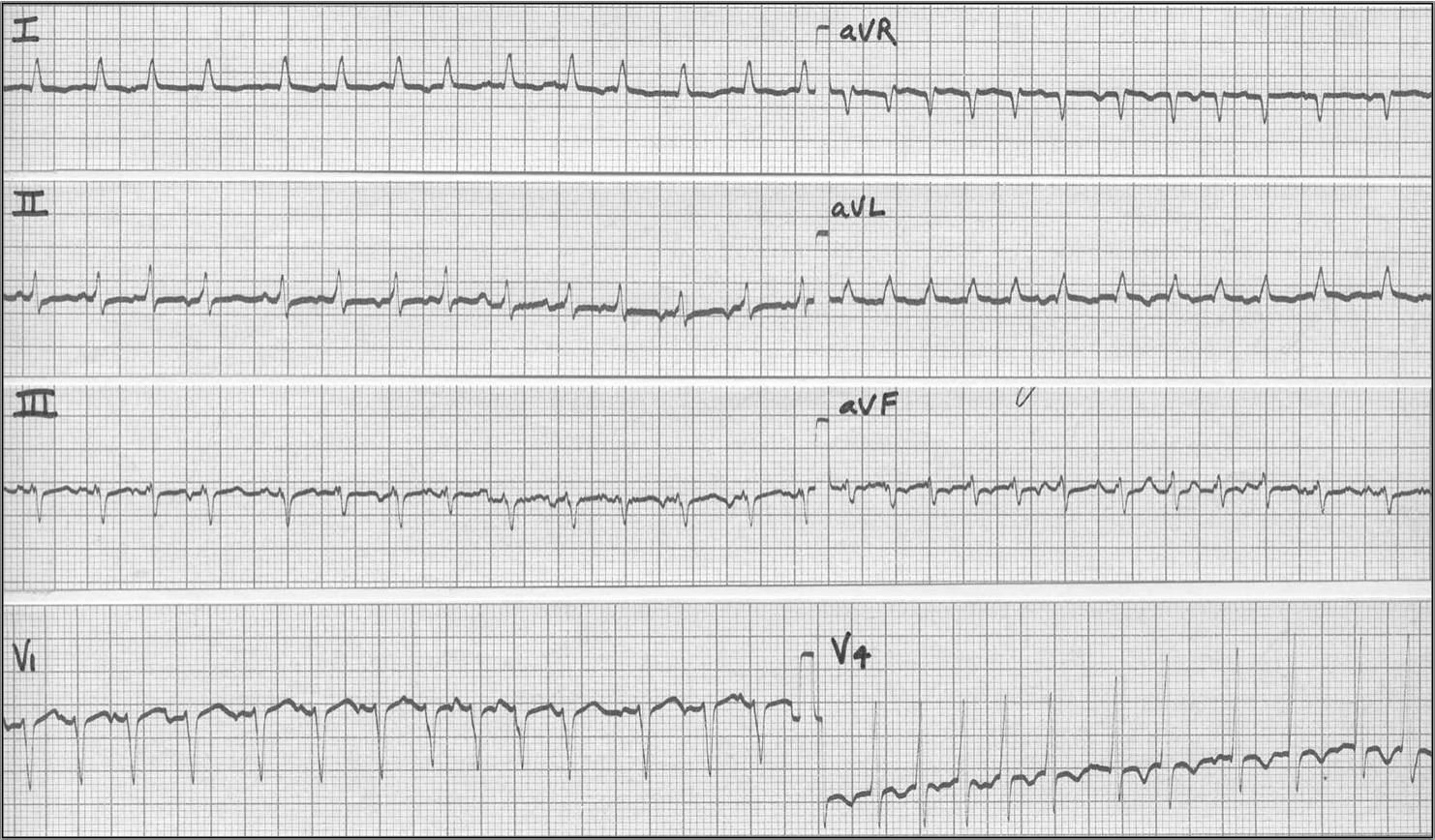
- A Very Subtle Atrial Tachycardia With 2:1 Block-KH
- Junctional Tachycardia With Exit Block: A Manifestation of Digitalis Intoxication-KH
- Atrial Tachycardia With 2:1 AV Block: A Manifestation of Digitalis Intoxication-KH
- Atrial Flutter with 2:1 AV block
- Atrial Flutter with 2:1 AV Block
- Atrial Fibrillation With Rapid Heart Rate Response and Left Ventricular Hypertrophy
- Atrial Flutter with 2:1 and 4:1 AV block
- Multifocal Atrial Tachycardia
- Onset of AV Nodal Reentrant Tachycardia
- RV PVC's
- Atrial flutter 2 to 1 in III and aVF
- Accelerated Junctional Rhythm
- Atrial flutter with mostly 2 to 1 AV block
Ventricular Rhythms and Tachycardias
- Ventricular Tachycardia With AV Dissociation, Captures, and Fusions
- Accelerated Ventricular Rhythm With Retrograde Atrial Capture and Echo Beats-KH
- Ventricular Tachycardia With Retrograde Wenckebach-KH
- Left Ventricular Tachycardia
- Ventricular Tachycardia
- Right Ventricular Tachycardia
- Supraventricular Tachycardia with RBBB
- Ventricular Fibrillation
- Torsades de pointes
- Ventricular escape rhythm in setting of Type II AV block
- VT strip w fusions and captures
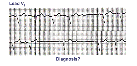
Mischief in the AV Junction (Blocks and Dissociation)
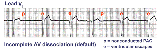
- AV Dissociation by Default
- AV Dissociation by Default
- AV Dissociation by Usurpation
- Isochronic Ventricular Rhythm
- 1st Degree AV Block
- 2nd Degree AV Block, Type I
- 2nd Degree AV Block, Type I, with Junctional Escapes
- LBBB and 2nd degree AV Block, Mobitz Type II
- Trifascicular Block: RBBB, LAFB, and Mobitz II 2nd Degree AV Block
- RBBB plus Mobitz II 2nd Degree AV Block
- LBBB plus Mobitz II 2nd Degree AV Block
- Mobitz II 2nd Degree AV Block With LBBB
- Incomplete AV Dissociation Due To 2nd Degree AV Block
- 2nd Degree AV Block, Type I With Escapes and Captures
- 3rd Degree AV Block Rx'ed With a Ventricular Pacemaker
- Complete AV Block, Junctional Escape Rhythm, and Ventriculophasic Sinus Arrhythmia
- 2nd Degree AV Block, Type I, With Accelerated Junctional Escapes and a Ladder Diagram
- ECG Of The Century: A Most Unusual 1st Degree AV Block
- ECG Of The Century - Part II: Dual AV Pathways
- Nonconducted And Conducted PAC's
- Two Wrongs Sometimes Make A Right
- Atrial Echos-KH
- Second Degree AV Block, Type I, With 3:2 Conduction Ratio-KH
- Second Degree AV Block,Type I, With Bradycardia-dependent RBBB -KH
- Supernormal Conduction: 2nd Degree AV Block With Rare Captures; Accelerated Ventricular Rhythm-KH
- 2nd Degree AV Block With Junctional Escapes And Captures-KH
- First Degree AV Block - Marquette-KH
- 2nd Degree AV Block, Type I (Wenckebach)-KH
- Complete AV Block (3rd Degree) with Junctional Rhythm-KH
- AV block high grade
Bundle Branch, Fascicular Blocks, and WPW
- Left Anterior Fascicular Block (LAFB)-KH
- LAFB: Frontal Plane Leads-KH
- Left Bundle Branch Block (LBBB)-KH
- LBBB: Precordial Leads-KH
- RBBB With Primary ST-T Wave Abnormalities-KH
- RBBB with Primary ST-T Abnormalities: Precordial Leads-KH
- Bifascicular Block: RBBB + LAFB
- Bifascicular Block: RBBB + LAFB-KH
- RBBB
- WPW Type Preexcitation
- Infero-posterior MI & RBBB-KH
- Infero-posterior MI & RBBB: Frontal Plane Leads + V1-KH
- Inferior MI and RBBB-KH
- Inferior & Anteroseptal MI + RBBB-KH
- Anteroseptal MI With RBBB: Precordial Leads-KH
- Atypical LBBB with Q Waves in Leads I and aVL-KH
- Atypical LBBB with Primary T Wave Abnormalities-KH
- Infero-posterior MI with RBBB-KH
- RBBB + LAFB = Bifascicular block-KH
- RBBB + LAFB: Bifascicular Block-KH
- Right Bundle Branch Block (RBBB)-KH
- WPW and Pseudo-inferior MI
- WPW with a Pseudo-inferior MI
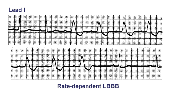
- Rate-dependent LBBB-KH
- Bradycardia-dependent LBBB With Carotid Sinus Massage-KH
- Left Anterior Fasicular Block: Frontal Plane Leads-KH
Artificial Pacemakers
- Ventricular Paced Rhythm With Retrograde Wenckebach-KH
- Ventricular Pacemaker Rhythm-KH
- Ventricular Pacemaker Rhythm: V1-3-KH
- Ventricular Pacemaker: Demand mode functioning-KH
- Atrial Pacemaker Rhythm
- AV Sequential Pacing
- AV Sequential Pacing
- Electronic Atrial Pacing
- AV Sequential Pacemaker
- Pacemaker Failure to Pace
- NSR with intermittent demand ventricular pacing
Myocardial Infarctions
- Anteroseptal MI: Fully Evolved-KH
- Anteroseptal MI, Fully Evolved: Precordial Leads-KH
- Extensive Anterior/Anterolateral MI: Recent-KH
- Extensive Anterior/Anterolateral MI: Precordial Leads-KH
- Acute Anterior MI-KH
- Infero-posterior MI-KH
- Inferior MI: Fully Evolved-KH
- Fully Evolved Inferior MI: Frontal Plane-KH
- Acute Inferoposterior MI-KH
- Postero-lateral MI: Fully Evolved-KH
- Postero-lateral MI: Precordial Leads-KH
- Diffuse Anterolateral T Wave Abnormalities-KH
- Infero-posterior MI & RBBB-KH
- Infero-posterior MI & RBBB: Frontal Plane Leads + V1-KH
- Inferior MI and RBBB-KH
- Inferior & Anteroseptal MI + RBBB-KH
- Anteroseptal MI With RBBB: Precordial Leads-KH
- Acute Inferoposterior MI with Right Ventricular MI
- True Posterior MI and Right Ventricular MI
- Old Infero-posterior MI-KH
- Old Inferior MI-KH
- Old Inferior MI, PVCs, and Atrial Fibrillation-KH
- Old Inferior MI-KH
- Atypical LBBB with Q Waves in Leads I and aVL-KH
- Atypical LBBB with Primary T Wave Abnormalities-KH
- Infero-posterior MI with RBBB-KH
- High Lateral Wall MI (seen in aVL)-KH
- Frontal Plane: Accelerated Junctional Rhythm and Inferior MI-KH
- Inferoposterior MI-KH
- Inf MI with RV MI and R sided ECG leads
- Acute inf STEMI
- Old inf MI
- Old interposterior MI
- Acute posterior MI in 15 lead ECG
- Inf MI with RV MI and R sided ECG leads
- Acute MI due to L main occlusion
- Inf MI with RBBB
- Anterior MI with RBBB
- Acute anterior STEMI with LBBB
- Old septal MI with LBBB
- Hyperacute T waves in inferior wall STEMI
Hypertrophies and Enlargements
- Left Atrial Abnormality & 1st degree AV Block-KH
- Left Atrial Abnormality & 1st Degree AV Block: Leads II and V1-KH
- Left Atrial Enlargement & Nonspecific ST-T Wave Abnormalities-KH
- Left Atrial Enlargement: Leads II and V1-KH
- Right Ventricular Hypertrophy (RVH) & Right Atrial Enlargement (RAE)-KH
- Right Axis Deviation & RAE (P Pulmonale): Leads I, II, III-KH
- Right Atrial Enlargement (RAE) & Right Ventricular Hypertrophy (RVH)-KH
- Bi-Atrial Enlargement with LVH
- RAE & RVH-KH
- Voltage Criteria for LVH
- LVH with "Strain"-KH
- LVH and Many PVCs-KH
- LVH & PVCs: Precordial Leads-KH
- LVH with limb-lead voltage criteria
- LVH: Limb Lead Criteria-KH
- LVH: Limb Lead Criteria-KH
- RVH due to atrial septal defect
- RVH with Right Axis Deviation
- RVH and LAE due to mitral stenosis
- RVH in 18 yr. old patient with pulmonary hypertension
- LVH: Strain pattern + Left Atrial Enlargement-KH
- LVH - Best seen in the frontal plane leads!-KH
- Severe RVH
- Left Atrial Enlargement-KH
ST-T and U Wave Abnormalities and Long QT
- Long QT with Giant Negative T waves
- Long QT Interval-KH
- Long QT Interval-KH
- Normal Variant: Early Repolarization-KH
- Normal Variant: Early Repolarization-KH
- ST Segment Depression-KH
- ST Segment Depression: Precordial Leads-KH
- Inferolateral ST Segment Elevation-KH
- ST Segment Elevation: Frontal Plane Leads-KH
- Long QT: An ECG Marker For Sudden Cardiac Death-KH
- Hyperkalemia and Old Inferior MI
- Advanced Hyperkalemia
- Giant TU Fusion Waves-KH
- Hypothermia: J-waves or Osborne Waves
- Hereditary LQTS
- Hyperkalemia T waves
- Brugada 12 lead ECG
- Brugada evolution due to TCA overdose
- Normal U waves
- TU fusion waves in CNS disease